The Medical student Review

  • Thread starter Thread starter جوري
  • Start date Start date
  • Replies Replies 123
  • Views Views 513K
Status
Not open for further replies.
:sl:

Sis I'm taking Organic Chemistry and Genetics, would that be a smart move to do at the same time? And will these notes help me in them?


:sl: sister

with all honesty organic chemistry was my downfall in undergrad, not even calculus I, and II were as bad as ochem -- sob7an Allah those who got it got it, but I wasn't one of them.. I had to drop it first time I took it and when I finally passed it, it was barely by the skin of my teeth, I was just so glad to have it behind me considering there were two courses of O.chem, and my happiness was short lived with one, on how I was to get by in the other..

my advise is to get a tutor early on, the first couple of chapters are deceptively simple but you can fall behind fast.. nothing on this thread pertains at all to O.chem.. maybe a few things on genetics, but undergrad is about understanding how things work together, graduate school is about their relevance to the real world, you can't get to one level without having completed the other as your knowledge base to build upon..

Now your experience might be completely different than mine -- I think if anything you should get out of this thread is to do at least just a little bit every day and not let it build up...genetics was fairly easy, it takes dedication so it is up to you.. the problem isn't with genetics it is with O.chem.. can you manage your time wisely or should you dedicate yourself to the difficult course get it behind you and take genetics later? only you are the best judge of your time and your ability..

and Allah swt knows best

:w:
 
a few skin conditions and later will go over dermatological pathology in common diseases of infants...

erythrasma_39806_lg.jpg


Erythrasma is a long-term bacterial infection that usually appears in the area between overlapping skin (skin folds).
Caused by a bacteria Corynebacterium minutissimum.
Erythrasma is more common in warm climates. in diabetics
appears link a ring worm infection but it isn't examine under woodslamp.. coral red because of coroporyphyrin III in the stratum corneum


fot3_peq.jpg


inverse Psoriasis--Inverse psoriasis is an unusual type of psoriasis that occurs in skin folds. These patches look different than other types of psoriasis. They are usually smooth, deep red, and glistening without any scale

1intertrigo3.jpg


intertrigo refers to an inflammation of the body folds. This is usually located in the inner thighs, armpits, and underside of the breasts or belly. It is a form of contact dermatitis.


22320084-1.jpg


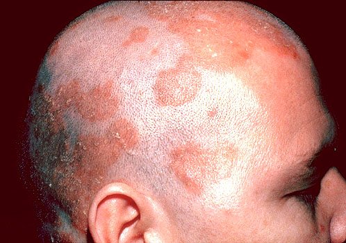
Seborrheic dermatitis is a papulosquamous disorder patterned on the sebum-rich areas of the scalp, face, and trunk. In addition to sebum, this dermatitis is linked to Malassezia, immunologic abnormalities.. when resistant to treatment think pt with undiagnosed HIV.
 
Last edited:
corresponding coronary arteries that supply regions of the heart
1- inferior wall st segment elevation in leads II, III, and AVF= right coronary artery
2- posterior wall (tall broad R waves in V1, V2, ST depression; and tall, upright T waves in V1, V2, V3) =posterior descending artery
3-Anteroseptal wall ( ST elevation from V1-V3) left anterior descending artery
4- Anterior wall (ST elevation V2-V4)= left anterior descending artery
5-Lateral wall ST elevation in I, AVL, V4, V6) circumflex artery

em_0028.gif

 
a Hodge bodge of review notes I know, but many are either high yields or areas that have caused me personal difficulty so for the sake of the greater good ...

a young woman presenting with nausea, diarrhea, abdominal cramps, vomiting for at least two weeks, her other hx is noncontributory, she does drink 'socially' has mild lower abdominal tenderness and these are her lab results

sodium 136/meq
potassium 3.0/meq
chloride 116/meq
bicarbonate 10/meq
BUN 16
cr 0.9m/dl
glucose 90
urinary sodium 12
urinary potassium 10
urinary chloride 110

the cause of her metabolic acidosis is?
diarrhea

because:

the patient has a non anionic gap metabolic acidosis
you calculate the anion gap as such :

(Na+ + K+) - (cl- +HCO3-) in this case gives us = 13 the normal value is (10-12)
anyhow,
the two most important factors of non-anionic gap metabolic acidosis are diarrhea where there is loss of bicarbonate in the stool and renal tubular acidosis (there are four) in which there is an inability to excrete sufficient amounts of hydrogen in the urine . the best way to differentiate between the two is to calculate the hydrogen ion excretion in the urine .. one expects hydrogen ion excretion in the urine to be high in diarrhea because the kidney attempts to get rid of excessive acid load -- and it will be low in RTA.
urinary hydrogen concentration is calculated as such: H+ = Cl- (K+ + Na+)
 
covering the topic of nephrology today:


figure out what kidney is failing?
______________

your approach:

renal failure has three main causes
pre-renal azotemia .. kidney not to function not because the problem is in the kidney rather in the circulation.

the second is intra-renal azotemia, or intrinsic renal disease--
and the third is post renal azotemia

problem with inflow to the kidney, or the outflow being blocked or can have disease within the kidney.

Now we start with pre-renal azotemia:
pre-renal many think of as dehydration, it is true in part, but it is a problem that is caused by poor perfusion, cause occur from dehydration and volume depeltion, or lost fluid from burn, third spacing during pancreatitis, or bad burn, kidneys aren't getting enough blood flow in, can have also cardiovascular disease, hypotensive because of poor cardiac output, not enough circulation to the kidney will be read as poor blood volume. inadequate blood to the kidney.

the same thing can happen if you have a situation of low albumin level leading to decreased oncotic pressure, like in nephrotic syndrome or cirrhosis, dec. intravscular volume because they run a low vascular volume. Kidney thinks it isn't getting enough blood.

the final main cause, is overt circulatory problem, like narrowing of renal artery, stenosis. Or medication induced vasconstriction, NSAIDS for instance, some of the blood flow is prostogandin dependent. cause the kidneys not to function and cr. goes up.

these are the causes of pre-renal. Not enough blood flow and not just because of dehydration.

why is pre-renal azotemia important. Important, conceived of as ischemic situation, if stays untreated, it will progress to where the kidneys necrose.

what you'll need to do is tell pre-renal from intra-renal because treatments are completely different.

________________

Next post renal, outflow problem

1- outflow of bladder can be obstructed, tumor in pelvis, scarring, strictures, BPH, back pressure goes back to kidney, causing obstruction of outflow..

or at level of ureters, stones, tumors, papillary necrosis, also putting back pressure unto the kidneys.
significance is back pressure on kidneys and damage

_______________

intra renal failure.. problem with the integrity of kidney, the most complicated of the three kinds..

intrinsicrenal, often acute tubular necrosis are many names due to kidneys not working.
how do you distinguish:
specify tests or lab data to interpret:

tests for post renal azotemia
first prostate exam, obstructed urethral outflow.
the best test however, check for residual urine in the bladder, have them urinate put catheter or scan with bladder scan to see how much urine is left after they urinate, typically they should only have 50cc or less

the best of all is an ultrasound, that looks at ureters, bladder.. which will detect hydronephrosis

now pre-renal azotemia

bunch of tests, the most widely used, is the ratio of BUN/CR
Cr level of 1
BUN 10
BUN more than 10 more than 30 it is a good indication of pre-renal
so that is a key labtest

the other more accurate
is measurement of urine sodium or fractional excretion of sodium

if kidneys are in a pre-renal situation , the kidneys say I am not getting enough blood, they think you're bleeding to death, so when they think that will, start retaining sodium to keep vascular volume, so urine sodium will be very low. short term treatment is fluids

urine sodium less than 10 is low

other things, same principal.. kidneys will also concentrate urine, high specific gravity and high urine osmolalaity, they are corroboratory findings.

typical case.
62 yr old for nausea and vomiting
his crt is 3.6 was normal 3 months ago, he also has orthostatic hypotension
BUN 82
urine sodium 6
specific gravity 1.028
BUN is more than 20:1
kidneys trying to replete vascular volume.. faced with this, this is pre-renal azotemia.
rx: fluids

_______________

intra-renal will see the opposite
BUN/CR is normal not elevated 10:1 or even less
will see instead of low urine sodium it will be high, because the kidneys are failing, they start leaking, so high levels of urine sodium
only place that falls down is pt who is on diuretic, so the test then loses value.
look for low BUN/CR
look for sodium spillage in urine
urine specific gravity and osmolality is low

you might also see red blood cell casts or granular casts, the kidney is falling apart and pieces of it coming apart, released into urine.

algorithm: approach to Azotemia

1- faced with high creatine level first thing you want to do is to exclude pre-renal azotemia :
a- what is the BUN/CR if >20:1
b-Urine Sodium <10
c-FeNA <1
d-OSM >300
f-specifc gravity > specific gravity rising by.001 for every 35 to 40 mosmol/kg increase in osmolality. Thus, a urine osmolality of 280 mosmol/kg (which is isosmotic to plasma) is usually associated with a specific gravity of 1.008 or 1.009. so higher than those values and now you know how to calculate 'normal'

sounds like pre-renal

if pre-renal is the case, then treat the cause, whether volume depletion, renal artery stenosis or heart failure..

also need to exclude post renal, using sonogram and post void residual urine, if see hydronephrosis then treat obstruction with stent etc
if no evidence or pre or post renal, then the problem is intra renal

BUN/CR not high
urine sodium that is high more than 20 or more than 30, FeNA that is greater than 1 and osm less than 300 and urine specific gravity is low since urine can't concentrate since kidneys aren't working properly.. that will tell you intrarenal ..

intrarenal failure there are many causes:
but they will either occur in the tubules and interstitum and the other occur in glomeruli which is glomerulonephritis ...

main categories:

first category and most important is ATN
acute tubular necrosis ..
occurs in different settings.. the tubules start falling apart, for instance in prolonged ischemia (shock kidney) hypotensive, labs look pre-renal but then kidney falls apart.. prolonged above conditions, clots of A fib, occluding renal artery...
2nd which are also common are toxins like
radiological contrast, drugs like amonoglycosides and amphotercin B, pigments like myoglobin from muscle breakdown

ATN occurs in phases, prodromal period of time
from beginning of insult to effect on kidney..
then when you have outright ATN, stop making urine, drop to nothing or 2cc an hr (oliguric) then there will be a diuretic phase of ATN
metformin with contrast are at a great risk..

another category is acute allergic interstitial nephritis, an allergy to a drug.. common drugs cephalosporins, beta lactams, methcillin hence we don't use it, sulfas, allopurinol
acute allergic nephritis, takes offending drugs, they'll get fever, rash, high eosinophil count, especially in the urine.
rx steroids short term
other tubulointersistial disorders
deposition disease, any number of chemicals can get into kidney, like hemoglobin is hemolyzed and out in circulation can deposit, myoglobin from rhabdomylisis, rx with high flow fluid to keep from crystalizing.
pts with Multiple myeloma crystalize Bence John's proteins
or crystalization of crystals, like oxalate, ethylene glycol antifreeze, part of mechanism of toxicity, is oxalate crystals depositing in kidney, also vitamin C in very big doses, can cause stones and crystalization.
crystalization of uric acid, for any disorder, like someone with lymphoma and it breaks off giving out uric acid, also in gout as well.
hypercalcemia, hyperparathyroid and crystallize calcium in the interstitum etc etc.
other causes of tubular interstitial disorders, infections like pyelonephritis, develop intrarenal, with white cell casts in urinary sediment.
others drugs and toxins, various analgesics NSAIDs, antibiotics.. outdated tetracycline.
variety of cancer deugs, cisplatin, mitomycin, methotrexate etc.
radiation nephritis, heavy metal poisoning...

glomeruldisorders

vasculitis type disorders (not very common) like wegner's granulamtosis
disorder with granulomatous dz in lung, paransal sinuses who also get lesions in the kidneys, intrarenal failure, high sodium in urine, cr high, problems with chronic sinusists and nasal congestion, maybe lesion in the lung as well.. test here is ANCA

Henoch shcnolen purpura, thrombotic thrombocytopenic purpura, htn etc.

other glomerular disorders, glomuerlonephritis, post streptococal glomerulonephritis.. get hypertensive and edema
do ASO titer to prove that the glomerulonephritis in someone who had a sore throat== post streptococoal glomerluonphritis

IGA nephrpathy, deposition of IgA following a viral illness...

chronic, like with collagen vascular dz. like SLE, also goodpasture syndrome
antibodies against basement membrane and lungs, will show up with intrarenal failure, and they will have lesions in lung and cough blood. order antibasement membrane antibodies

another Alport renal failure with hearing failure.
___________________________________________________


Now, how will we decide who has what...

glomerulonephritis

high urine sodium >20
High fractional excretion of sodium >1
BUN/CR suggesting not pre-renal
urinalysis will be key
hematurea and RBC casts = glomerulonephritis
most definitive tests to prove Glomerulonephritis is a kidney biopsy

things that go along with glomerulonephritis is nephrotic syndrome, also occurs in vascuilitis..
1- massive proteinurea massive 3.5 g 4+ dipstick positive and more
they lose intervascular oncotic pressure and develope edema, will run albumin down and hyperlipdemia

best test for nephrotic syndrome is a 24hr urine protein
best test to determine the type of glomerulonphritis causing this nephrotic syndrome is a biopsy.

some nephrotic syndrome are treated with prednisone

best first test (initial test) in a pt. with high urine sodium and indicators of intra renal failure.. is urinalysis!!!


infection, drug hypersensitivity, glomerulopnephritis, nephrotic syndrom, meyloma, uric acid (gout) and myoglobinurea.

what do we see in each of these?

infection= WBC and bacteria
pyelonphritis WBC casts
Drugs/ eosinophillia
glomuerlonephritis = RBC casts
Nephrotic syndrome = Hyperlipedmia, heavy protenurea
Multiple myeloma _ proteinurea
uric acid = uric acid crystals
Myoglobinurea = High CK levels and dipstick positive test for blood but no blood cells.


a different way
pyuria = infection
RBC casts = glomerulonephritis test with kidney biopsy
blood urine dipstick but not micro = myoglobinurea
crystals in uine = gout
eosinophils = allergic drug reactions
massive proteinurea = nephrotic syndrome
__________________________________________

cases:

62 yr old nausea vomiting, creatinine is 3.6 was normal 2 months ago, takes no meds
has large prostate, pulse is high, BUN is 72, urine specific gravity is 1.023
urine sodium = 9
all else is WNL

best initial rx..
likely cause.. he has a large prostate gland, potential outflow obstruction
tempted to get sono to measure post void.
his other things.. BUN 72 and cr 3.2 ratio of 20
concentrated urine
urine sodium of 9
all point to pre-renal azotemia
best initial rx is fluids
_____________________

case II
62 yr old nausea vomiting. CR 3.6, taking NSAIDS
labs
urine specific gravity 1.004 dilute
BUN 41/ 3.6 almost 12:1 ratio
urine sodium = 42

renal problem spilling sodium, can't dilute urine even though dehydrated, he has renal disease, with many dx to consider..

what is next? do sonogram to exclude post renal azotemia, then go back and say renal dz and work it up.

________________________

boy with high BP, edema, urinalysis, proteinurea and RBC casts..
young boy with glomerulonephritis.. blood test to do is ASO titer.

__________________________________

ESRD

end stage renal disease
renal failure that is incompatible with survival..
need dialysis or kidney transplant

how does it show up?

chronic findings and acute findings
important points, need for dialysis isn't determined by creatinine

hypocalcemia
kidneys are supposed to make 1.25 hydroxy vitmin D, because they are not working, you don't have this chemical so don't absorb calcium in GI tract, thus your body makes more PTH to absorb calcium from bone, causing bones to become osteopenic. renal failure need their calcium and Vit D supplementation

people get hypermagnesemia because they can't secrete it.

also have accelerated HTN through renin angiotensin system, very difficult to control, combo of accelerated HTN and high lipids, causes death.

always anemia, since kidneys make erythropoeitin, pts on dialysis, will usually receive erythropoietin but anemia is concomitant to ESRD

impaired immune functions, as well presumably because of toxins not excreted get pruritic.

all of these things are common, become osteopenic and break hip, acceprated lipids and HTN and get MI.

Hyperkalemia, potassium high enough you'll die
potassium is too high from changes it makes on EKG, focus on T waves..
T waves are so sharp and pointed, you don't want to sit on them:

MMPE_12END_156_02_eps.gif


hyperkalemia, that is an acute manifestation that potentially needs dialysis..

metabolic acidosis, in non working kidneys, is severe enough fatal needs dialysis..

fluid overload to the point of pulmonary edema, if kidney can't urinate then dialysis

pericarditis, build up in pericardial sac needs dialysis

pts with high BUN, obtunded, encephalopathic, comtaosed needs dialysis.

renal dialysis, in blood, or direct connection in the blood, venous shunt, or pertoneal, fluid swish around and come back through cath.

________________________

62 yr old pt on dialysis, returns from 4 days trip, feels very poorly (pts need dialysis every two days) he has nausea, vomiting and SOB, he has tall peaked T waves. Cr. 5.2
Hco3- is 16
metabolic acidosis
best rx is dialysis immediately..

__________________

quick recap with key words:
Eosinophils in urine = drug reaction
RBC casts= glomerulonephritis
Heavy proteinurea= nephrotic syndrome
high triglycerides= nephrotic syndrome
Radiological contrast = ATN
Urine sodium < 10 pre-renal (kidney trying to retin its plasma volume)
BUN/CR > 20 = pre-renal
 
Last edited:
Part II nephro.

Now with electrolyte disorders.. very important...
follow this recipe and you can't lose :X


let's start with hyponatremia which is the most common:

SX that folks have, when it is mild and gradual, pts are asymptomatic, sodium falls slowly, with severe, they gets headaches and confusion, and abnormal mental status changes, and at an extreme that is rapid, they can develop CNS coma and seizures ..

causes of hyponatremia, or pseudo hyponatremia
in couple of situations, sodium is low when the blood is hyper-osmolar, what is ahppening the patient is hyperglycemic, sugar is high and as an artifacts of balancing fluids, the sodium falls... the relationship for every 100 mg glucose goes up the sodium falls 1.6mg

pseudo hyponatremia with normal osmolality, artifcat with high triglycerides or hyper-proteinemia like with multiple myeloma

and there is real hyponatremia

so another way to look at it, is you can have hypeonatremia with high osmolality which is seen in hyperglycemia
you can have hyponatremia with normal osmolality which can be seen with lipids or proteins .
or you can have hyponatremia with low osmolality and that can be seen with multiple causes..

____________________________________

high and normal is a pseudo..

so first step to determine hyponatremia is to look at the osmolality

going to continue this review tomorrow insha'Allah since I am falling asleep...:-[

____________________________________________________________

picking up from where we left off:

low osmolality multiple causes real hyponatremia..
first step is to determine the osmolality to see if psuedo or real..
send blood plasma but sometimes you have to figure out by calculating
formula: 2xserum sodium + glucose/18 + bun/2.8 = osmolality
or better easier, 2 x sodium + 10 is glucose is normal
normal osmolality is between 280~300

_____________________

hyponatremia with high osmolality.. there is a new solute in high concentration usually glucose, causes fluid shift.
1.6mg% for every 100% rise in glucose
ex. if glucose is 342 and sodium is 126, glucose account for 3.2 sodium drop..
how much higher than normal is glucose of 342? that is 200 units higher than it should be, so for every 100 unit, the sodium drops 1.6
200 points higher, 200x1.6 =
adjusted sodium is 129 so doesn't account for all the drop in sodium thought..

another example
glucose 342
sodium 131

does the glucose explain hypontremia?
400-100 = 300 units higher
300x1.6= 4.8
4.8 + 131 = normal sodium

____________________

now hyponatremia with normal osmolality
coming from proteins and lipids..

certain amount of blood is made of protein and lipid..
conc. of sodium
in abnormal situation, the protein and lipid is higher leaving less space for water and sodium
the amount of sodium is normal but less of it per liter of blood.

patient presents with weakness.
sodium 128
glucose 142
serum osmolality 282
total protein 7.2

what test will show cause of hyponatremia? lipids
normal osmolality hyponatremia..

____________________________

real hyponatremia, measure extra-cellular fluid volume
divide into multiple causes of low osmolality

determine volume status
if have high volume status, then edematous condition

ecf is low
fluid loss, from diuretics, kidney, sweat, burns, etc.

if ecf is normal euvolemic..
multiple causes like
SIADH (ecf normal category)
if high hypervolemic
if low hypovolemic
if euvolemic there aremultiple causes.

low osmolality with hypovolemia, volume depeltion either renal or extra-renal, going to tell by urine sodium.
burn patients or marathoon runners.
or get volue depletion from losing fluid from kidneys

hypoaldosteronism, glucocorticoids deficiency

the most important of the three conditions, the one that does it with a normal volume..

common cause is SIADH
other causes are pathological water intoxication, psychogenic polydipsia, low osmolality hyponatremia but will retain normal volume
hypothyroidism

the most important thought is SIADH
the urine osmolality is abnromally high in face of hyponatremia and low serum osmolality-- if blood is hyponatremic diluted, appear to have extra water in the blood, the kidneys ought to protect me, pee out water so that the sodium level can come back up? expect to have dilute urine to get rid of dilute water in blood.. but here the antidiuretic hormone is abnrmal, actually excretes very concentrated urine high urine osmolality in urine with low osmolality in blood, which should be fixed.. but it is because I have an inappropriate ADH causing me to retain fluid and get rid of sodium ..
high urine osmolaity when serum osmolality is low.. need to also look for renal or thyroid function or drug problem too.

SIADH causes are lung disease, can cause brain to secrete inappoporiate amounts of ADH, concentrate urine and dilute the blood. any brain disease, tumor, subarachnoid hemorrhage, small cell lung cancer, or drugs, like TCA, MAOi, fluxoteine, carbamezapine, narcotics, phenothiazines, clofibrate, vincrsine, cyclophosaphamide, vinblastine.

drugs that increase ADH from brain or on kidney, oxytocin in labor acts like ADH.

SIADH dx. serum hyponatremic, low osmolality, and urine ought to have low osmolality but instead inappropriately high.

rx: either reverse the cause, but you can't always do that,if you can't reverse the cause, restrict fluid, or give furosamide with normal saline..

or two drugs that decrease the action of SIADH on the kidney
demclocycline and lithium
emerency rx for serious hyponatremia as convulsion and seziures is normal saline.

someone comes in with hyponatremia
measure serum osmolality and see if high low or normal.

figure out volume status.
if high, edematous conditions, heart, liver failure
if ecf is low, they are fluid depelted, measure urine sodium, if low they are losing from skin or GI
if high then probably losing it from the kidneys

if fluid volume is normal,
then SIADH or previousely discusses..

cases:

82 yer old vomitting and confusions, takes thyroxine, fluoxetine, glucose 342, has pneumonia

he is vomitting can cause dehydration
low ecf volume hyponatremia
taking thyroid med can cause SIADH like syndrome
as well fluoxetine and chlorpropmide
glucose is high running glucose level down
how to determine?

need osmolality and volume status as well urine sodium to figure out
if efc is normal then check urine sodium
_________________________________________

again

approach to hyponatremia


key points..
Determine serum osmolality high and normal are artifacts of glucose lipids and proteins
with low osmolality determine the ECF volume
High ECF edematous state
low, renal or extra renal depending on whether the sodium is greater than 10 or less than 10
normal ECF then SIADH, drugs, thyroid, cancer etc

_______________________________________________

Next topic is hypernatremia which is the least common of these metabolic disorders:

cause of hypernatremia
people typically dehydtrated, losing fluid more than sodium, thus sodium level rises, occurs through insensible water loss, people with bad burns, also common from GI tract in diarrhea, and become hypernatremic that drive to drink more when you go alot..

another cause is diabetes insipidus, no ADH action, ADH either not made or can't work on kidney, so either a tumor in pituitary or such or nephrogenic, ADH is made but doesn't work on kidney, due to lithium or kidney failure, spill water in urine like crazy..
lethargy, sezures and coma like hyponatremia

rx is fluids, treat the dehydration, think of diabetes inspidus like you'd think of diarrhea..
correct the problem slowly, as to not cause serious central myelnosis

if can't correct this problem right away think diabetes inspidus..
do water restriction test

with passage of time measure urine volume and urine concetration as we restrict their fluid intake..
in a normal person. the urine volume will be less with restriction and the urine comes out should be yellower and more concetrated.
if you have diabetes insipdus, you don't have any ADH, you'll mak urine at same volume, if give antidiuretic at that stage and are unable to respond because of kidneys it will keep right on then you can make dx of nephrogenic diabetes inspidus.. on the other hand if given antiiduretic hormone and it become normal then you've responded to it, you had a CNS lesion that prevented you from making ADH.

rx correct the underlying problem, if tumor or lithium, or replace antidiuretic by given internasal DDAVP
if nephrogenic because of kidney defect, you can give thiazide diuretic causing sodium depeltion helping keep water or NSAIDS impair ability of concentrating ability, neither of those methods are really all that great.

__________________________

now low potassium problm hypokalemia

common causes, GI loss, diarrhea.. various tube drainages in the bowel, medications, diuretics, people on lasix and furosamide, beta agonists like albuterol cause calcium to enter into cells, decreasing k+ in circulation.
Insulin, and drive sugar from circulation into cells potassium goes with it.
then there are rare causes

hypoaldosteronism (conn's syndrome) primary hypoaldosertonism, cushing's syndrome, bartter's syndrome, can't absorb sodium chloride, lose potassium along the way.

liquiorice, not common but has mieralcorticoid effect. Glyceric acid.. chemical in liquiorice with mineralcorticoid effect.


Hypokalemia

73 with COPD and CHF, has diarrhea for three days, he is on lasix, furosamide, and multiple albuterol rx in the ER

multiple causes of hypokelamia

diarrhea in stool
lasix in urine
multiple albuterol (potassium to go into cells

mild is specific t wave changes, severe can cause cardiac toxicity as well potentiate digoxin effects, because of hypokalemia

rx replace the potassium, orally, best if possible..
but in hospital pts at times given IV 10meq/hr

too much potassium can cause cardiac conduction defects, this is how you execute people in prison, you cause heart block and asystole
____________________________________

hyperkalemia
increased intake of potassium, error in hospital, or meds that causes hyperkelamia, like potassium sparing diuretic, ace inhibitor, potassium artificat from venipuncture...
the tourniquet and blood sample hemolyzed and caused potassium to go extracellularly..

also in situations where potassium moves from cells into blood, like familial periodic parlaysis, myoglobuniurea, renal failure, drugs. hypoaldosteronemia

adrenal inssufiency ..

tall pointed peaked t waves..
pt on ace inhibitor or on NSAIDS or spirnolactone or others and potassium is high, specify treatment

67 y/s with DM on glyburide, and ace inhibitr
cr 4
and potassium 6.6
number of reasons
ace inhibitor
because cr is rising renal failure can't excrete potassium
how will you fix it?

in stages:

first stage if you get an EKG changes protect the heart stabilize cardiac membrance
so give calcium chloride or gluconate infusion.

next lower the potassium level by driving potassium out of circulation by giving glucose and insulin 50% dextrose and insulin or give doses sodium bicarbonate create a situation of alkalosis, potassium will go into cells
or get rid of potassium from body keyexylate
or if in renal failure then dialyze them

number one with EKG changes protect the heart give calcium
number two lower serum potassium
if no EKG changes then lower potassium and get it out of the body

_________________________________________________________

Now for acid base disorders (RTA's) acidosis and alkalosis.
have a systematic approach to it all...interpret blood gases...

alkalosis= PH is high
acidosis = PH is low

first step is to determine acidosis or alkalosis..
7.4 is normal
less than 7.37 acidosis if more than 7.4 alkalosis
first level decision

now decide whether respiratory or metabolic acidosis or alkalosis..

arterial blood gases..
get PH, will get PCO2 normal is 35-45 and dependent on breathing rate, the faster the lower the PCO2, the slower the Higher

bicarbonate normal is (20-28)

normal mid twenties.. bicarbonate is a metabolic buffer
and changes develop slowely..
if bicarb is trying to fix acidosis, it will happen over days as opposed to PCo2 in a matter of minutes

O2 and PO2 level which varies by altitutde, and saturation which should be over 90

let's start with respiratory alkalosis

caused by a bunch of things
hyperventillation, rapid acute fast breathing..
respiratory alkalosis will drive PCO2 now, various causes, can be caused by anxiety, acute pain, panic attack, and if this happens, PCO2 will go down but there is no other problem

other acute prbs, like Pulmonary embolism and pneumonia, also early asa toxicity, breathe fast, breathe down their PCO2..

no time for metabolic compensation... will see high PH, and PCO2 that is low because of fast breathing, and HCO3 didn't have time to change so it will be normal..

24 year old female, feeling suffocating and tighteness in chest, RR 36
get blood gases, find out PH 7.52 alkalotic
O2 is 99%
PCO2 is low and HCO3-

thus respiratory lkalosis

__________________

another 24 year old everything the same
PH 7.52
however her PCO2 25 and her O2 sat is 76% which is low
HCO3- 27

there is a respiratory problem

ON BCP and had a PE
she is hypoxemic

___________________

metabolic alkalosis
the problem is you are gaining an alkali bicrbonate or you are losing an acid
how does it happen?
lose acid with prolonged vomiting or other GI loss, like NG tube, or diuretics or rather than losing acid, gain HCO3- people who use sodium bicarbonate pills (antacids) calcium bicarbonate for antiacids build up of bicarb, or H2 blockers or proton pump inhibitors..
it is a slow chronic process, losing acid, or gaining bicarb gradually.. as you start to become alakaline, the body will breathe slower so that PH falls from alkaline level to closer to normal..

increasing PH almost back to normal..

typical case..
67 yr with HTN on HCTZ complains of fatigue, her exam shows dehydration with orthostatic, hypokalmic, hypnatremic, hypchloremic... PH 7.42 (a little alkalotic)
PCO2 is high and bicarbonate is high

alkaline, because she has high base in her blood, metabolic alkalosis. body compensating by breathing slow.

________________________

respiratory acidosis, hypoventillation.. could be acute or chronic.. an inability to blow of PCO2
COPD, can't exhale all the air, will build up PCO2 in blood..
breathing poorly become acidotic.. kidneys retain alakli (hco3)
acute respiratry acidosis .. inability to breathe at normal level, taking narcotics that lower respiratory rate, or ashtma..
on chronic basis.. chronically elevated PCo2 from Obesity, or COPD

ABG
dec PH
high PCO2
if acute the bicarbonate won't have time to rise..

typical case
27 yr unconscious
rr is 7
pupils is pinpoint
ph 7.28
O2 is low
PCO2 is high (not breathing)
no time for bicarb compensation thus it is normal

72 yr with COPD getting worst, getting more SOB
ABG
PH 7.37
O2 sat 90%
PCO2 is very high
HCO3- is also high

acidotic because she is breathing ineffectively, in attempt to crrect HCO3 tries to correct it

________________________________

question on the same ABG
Ph. 7.37
O2 sat 90%
pCO2 56 (high)
HCO3- (34) high
metabolic alkalosis (HCO3-) high with high PCO2 as compensation?

how do you know that the alkali isn't the primary thing?
because PH is not alkalotic it is acidotic.. without over comepnsating

______________________________

metabolic acidosis the most complicated...

causes a relative increase in the quantity of acid, due to any of the following, addition of acid, inability to excrete acid, loss of base HCO3-
mechanism in blood gases
low PH decreases b/c acidosis
bicarbonate dec because less base
respiratory rate will increase to blow of PCO2 to increase PH back to normal..
sudden acidosis, the respiratory rate will kick in right away

17 yr old with DM and not taking his insulin presents with altered mental status.. PCO2 is low and Bicarb is low, metabolic acidosis because the bicarb is low, and as comp. body has been breathing fast to blow off PCO2

first step in sprting metabolic acidosis is it?
metabolic with high anion gap, or normal anion gap, called non anion gap..


what is an anion gap?
it involves
adding the postive cations and subtracting from them the two negative anions as such

[NA+ +K+] - [cl- + HCO3-]

normal anion gap is 11+/- 3 i.e between 8-14

high anion gap acidosis higher than above number means another organic acid has been added to the body.. normal anion gap acidosis, either excess HCL- or loss of bicarbonate...

high anion gap acidosis a few causes
over production or under secretion of acid, chloride levels unaffected

four causes
1- ketoacidosis ( alcoholic ketoacidosis, diabetic ketoacidosis, starvation
lactic acidosis
renal failure
or intoxication with any number of different intoxicants
ethylene glycol, methanol, salicilates

la mud pie
l lactic acidosis
a aspirin
m methanol
u uremia
D DKA
P propylene glycol
I isopropyl alchol
e ethlyene glycol

normal anion gap acidosis

they are either losing HCO3- or can't excrete acid..
common causes of these are diarrhea, loss of bicarbonate, or RTA inability to excrete acid in the urine___________________

normal anion gap (RTA)

they all have one thing in common, inability to acidify the urine..
renal tubular acidosis kidneys can't do that.. less acidic urine or overtly alkaline which is wrong...

type I RTA occurs in distal tubule of the kidney, you can't acidify urine, your urine PH will be high 5.6 or higher
seen in kidney stones, amphotercin B toxicity, lithium tox, sickle cell disease. give acid challenge test.. expect that acid spills in kidney and they should acidify urine, but they can't.. the rx is to neutralize the urine by giving bicarbonate.

_____________

RTA type II which occurs in proximal renal tubules and the problem here is inability to absorob HCO3- until the levels becomes very low, eventually in the distal tubule some bicarbonate will be salavged..so again inability to acidify the urine
occurs, in myeloma, fanconi syndrome, wilson's disease
RTA II inability to absorb bicarbonate..
bicarbonate is lost in urine even in face acidic blood, and the treatment here is volume restriction , the kidneys will absorb a little more

Type IV hypoaldosteronism, adrenal defienciy, some diabetes..
the presentation is key, presents with hyperkalemia..
restict salt, and even in restricing salt, the sodium is lost, it is treated with fludrocortisone.

_______________________

so let's do chart

RTA type I

defect: Distal tubule cannot excrete acid.
Results: urine is alkaline even when blood is acidic
K+: is low
test is acid load test -- urine remains alkaline
RX: oral bicarbonate
________________________
RTA type II
defect: proximal tubule can't absorb bicarbonate at normal urine volumes
Results: urine remains alkaline except at very low urine flow volume
K+= low
Test: do a bicarbonate load test renal tubules don't absorb alkali and the urine remians alkaline
RX: volume restriction
_________________________
RTA IV
defect: renal or aldosterone deficiency
Results: in high urine sodium -- aldosterone usually causes you to retain sodium and lose potassium, and when you have an aldosterone defcicney you'll lose sodium and retain potassium. increase in sodium excretion in the kidney exchanges with potassium and hydrogen -- thus you build up high hydrogen ion -- that is how you get acid in RTA type IV
K+: high
Test: salt restriction test.. the urine sodium remains high, spilling sodium in urine
RX: replace mineralcorticoid with fludrocortisone..

___________________________________________


Last edited:
An interesting case of a 28 yr old woman who presents for eval of oligmenorrhea and infertility. P.E reveals acne, hirsuitim, and mild clitromegaly. Lab tests reveals mild hyponatremia, and normal levels of 17-ketosteroids and plasma dehydropiandrosterone levels and no change in aldosterone levels. The Pt is diagnosed with a defect in CYP21A2.. What best explains the late onset of her disease?

and the answer is an incomplete 21 hydroxylase defect.
The Patient described has disease resulting from partial dysfunction of CYP21A2 (21-hyroxylase P450C21). defects in CYP21A2 are the most common cause of CAH. There are two copies of each gene in the human genome, one derived from the mother and the other from the father. A complete deficiency of a gene product therefore, requires inactivating mutations in both copies of the gene in question. CAH evident in infancy thus requires that both genes for an enzyme required for adrenal steroid hormone synthesis be inactivated- Adult-onset CAH, however is most commonly the result of an inactivating mutation in one copy of a steroid synthesis gene. The remaining copy is able to code for sufficient enzyme to prevent catastrophic infantile presentation of CAH. In the case described, the patient's single remaining copy of CYP21A2 was able to synthesize sufficient aldosterone precursors to prevent extreme salt wasting and was able to prevent and extreme accumulation of steroid precursors, thereby preventing virilization in infancy. She does however, overproduce androgenic adrenal hormones on ACTH stimulation, and this is likely the cause of her oligmenorrhea, infertility, hisruitism and acne.


Now to look at other similar defects in the same pathway. A complete 3-hydroxysteroid dehydrogenase defect, would cause defects in aldosterone and cortisol and overproduction of adrenal androgens. Clinically male pts. would present with ambiguous genitalia and female pts with virilized genitalia. Hypertension isn't a feature of this disease..

11-hydroxylase (CYP11B1) deficneyc causes hypertension through accumulation of mineralcorticoid 11-deoxycorticosterone.
 
70 year old woman with a long history of DM brought to the ER with a sudden onset of weakness on the right side of the body. She has a 20 pack year hx of smoking and is currently taking metformin and hormone replacement therapy. She is oriented to time and place and person, and seems to answer all questions appropriately. She was able to dress herself before showing up to the ER. Her vitals are BP of 140/90 mmHg and PR of 85/min. P.E confirms loss of strength in the right upper body and lower extremeties. There is a right sided facial weakness as well. The visual field is intact. A CT scan of the head is ordered. Occlusion of which vessel is the most likely cause of her SX?

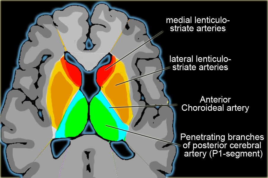
and the answer is penetrating arterioles of the posterior limb of the internal capsule. Lacunar infarcts are small (<0.5cm) focal areas of parenchymal loss found in the basal ganglia, anterior and posterior limb of the internal capsule, pone and occasionally cerberal white matter. The five classic syndromes include pure motor hemiparesis, pure sensory stroke, ataxic hemiparesis, and clumsy hand dysarthria. They are presumed to be ischemic in origin and due to occlusion of the pentrating arterioles aforementioned structures. Lacunar infarcs are particularly frequent in persons suffering from hypertension or DM. The most common type is a pure motor lacunar infarct most often occur in the genu or posterior limb of the internal capsule (Aphasia, Agnosia, Hemianopsia, neglect etc) pure motor infarcts most often occur in the genu of posterior limb of the internal capsule where the descending corticospial and corticobulbar tracts are located. Infarction of the anterior limb of the internal capsule, most often results in ataxic hemiparesis where both the pyramidal (weakness) and frontocebellar (ataxia) are located.

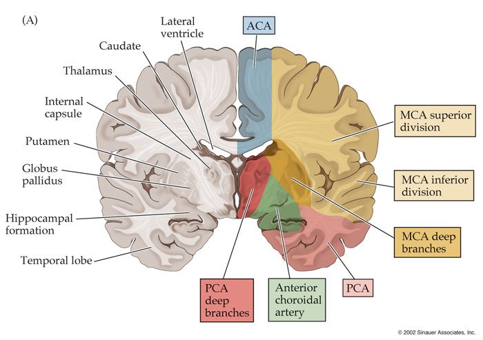
Now the other major arteries like Anterior cerberal artery occlusion presents with contralteral weakness and cortical sensoty loss in the contralteral leg without weakness in the arm. Urinary incontenance, confusion and behavioral distrubances are likely.

The Middle cereberal artery occlusion presents with contralteral hemiplegia, hemisensory loss, aphasias and homonymous hemianopia. our pt here has intact visual fields and not suffering aphasia.
Posterior cerberal artery occlusion presents with contralteral homonymous hemianopia visual hallucination and agnosias.

vertebral artery occlusion may not cause clinically noticeable sx because of good collateral circulation when sx occur they are similar to basialar artery occlusion.

490b6c1148232Territorybasalekernen-1.jpg


thmb_48e6c40d05324VD%20thal-infarct.jpg


Lacunar infarcts

CerArtDistBlum1-1.jpg


self explanatory...
 
I'll be writing the step up to USMLE step 3 on here.. will save most of you a good $40 insha'Allah..

we'll start with cardio ..

little factoids first..

the left anterior descending is the most common site of coronary artery occlusion
cor_art.gif


coronary arteries fill during diastole. while systemic arteries fill during systole. conditions or drugs that reduce diastolic filling decrease coronary perfusion during a given period of time.

As heart rate increases, the time available for diastolic filling decreases.
During exercise, cardiac output initially increases due to increased stroke volume and then due to increased heart rate.


  • cardiac output is dependent on the rate of contraction. I.E heart rate, and the volume of blood forced out of the left ventricle per contraction. i.e stroke volume.
  • heart rate is the number of contraction per unit time and is expressed as beats per minute.
  • stroke volume is the change in volume from immediately before a contraction to the completion of a contraction. SV = (end diastolic volume) - (end systolic volume)
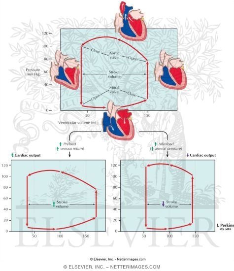
  • dependent on contractility i.e force of heart's contraction, preload i.e the amount of stretching force on cardiac muscle fibers at the end of diastole, and after load, i.e the vascular resistance which ventricles must overcome to produce outflow.
  • increases with catecholamine release e.g epinephrine, increased intracellular calcium, decreased intravscular sodium, digoxin use, and stressful events. e.g anxiety, exercise
decreases with beta blockers, heart failure, hypoxia with acidosis.
___________________________________

Vascular pressures
_______________________________

  • systolic blood pressure is the maximum vascular pressure expereinced during heart contraction, diastolic blood pressure. DBP is the baseline vascular pressure between contractions.
  • pulse pressure is the increase in blood pressure attributed to cardiac outflow during contraction
  • pulse pressure = (SBP)-(DBP)
  • mean arterial pressure (MAP) is the average BP considering the unequal amounts of time spent in systole and siastole
  • MAP = (DBP) + (1/3 X SBP)
_________________________________________
heartantmajorvessels.jpg


Aorta gives rise to the left and right coronary artery.

Aorta >>>>>>left coronary artery>>>>>>>left anterior descending branch >>>>>septal branch supplies anterior wall of left ventricle, the septal branch supplies anterior 2/3 of intraventricular septum.
Also from the left coronary we get the circumflex branch which supplies the left atrium lateral wall of left ventricle, posterior wall of left ventricle.

now from the AORTA TO THE right coronary we get three branches off the right coronary artery (the posterior descending branch which supplies the inferior wall of left ventricle, and posterior 1/3 of intraventrciular septum, the marginal branch supplies the right atrium, right ventricle, and the SA, AV node branches.. very important to pay attention to that infarction to the right coronary will spell disaster to the SA and AV nodes.

now the physiology of heart contraction.
Key principles


  • increasing the end diastolic ventricular volume causes an increaased stretch on cardiac muscle fibers, this leads to an increase in the force of contraction i.e the frank starling relationships
  • the end systolic volume and pressure generated by the ventricles are dependent on afterload
increased contractility i.e force of contraction independent of preload and afterload leads to increased muscle fiber tension during isometric contraction
_____________________________
 
49680550x0475-1.jpg


EKG
P wave, normal atrial depolarization
PR interval: conduction through the arterioventricular node (<0.2 sec)
QRS interval: ventricular depolarization (<).12 sec)
ST interval: isoelectric ventricular contraction
T wave: ventricular repolarization
U wave: relative hypokalemia

40221-1.png

___________________

Normal cholesterol function
Cholesterols and triglycerides are carried by lipoproteins
increased low density lipoprotein (LDL) levels lead to an increased coronary artery disease (CAD) risk
increased HDL is protective
Increased LDL and decreased HDL result from a diet high in fatty foods, tobacco and obesity, alcohol use, DM, and certain meds like OCP and diuretics.

Pre-operative cardiac risk assessment:
for surgical pt estimating the liklihood of an undesired cardiac event occuring as a result of surgery and anesthesia

  • considers measurable cardiac function, pre-existing cardiac dz, age and important co-morbidities
  • young healthy pts. maybe cleared for surgery with a normal EKG
  • older pts with existing co-morbidities require and extensive workup by a cardiologis
  • factors suggesting and increased risk of an adverse cardiac event
  • age >70
  • pulmonary function, forced expiratory volume in 1 sec/functional vital capacity (FEV1/FVC) <70% of expected. Partial pressure of carbon dioxide (pCO2) >45 mmHG, pulmonary edema.
  • cardiac dz: MI within the last 30 days, poorly controlled nonsinus arrhythmia, pathologic Q waves on the preoperative ECG, severe valvular dz., decompensated heart failure with poor ejection fraction.
  • Renal insufficiency: creatinine (CR) > 2.0 or a 50% increase from baseline
  • surgery type: cardiac/vascular surgery or anticipated high blood loss
  • higher risk pts should have their cardiac function optimized prior to elective surgery and should be made aware of the risks is the surgery is emergent
  • perioperative B blockers and postoperative noninvasive cardiac monitoring are recommended for pts determined to be at an increased cardiac risk.
  • ______________________
Invasive cardiac monitoring

  • Arterial line: constant access to artery, e.g radial, femoral, axillary, brachial, dorsalis pedis) that allows accurate measurement of arterial BP and allows easy access to arterial blood for blood gas measurments
  • pulmonary artery cath I.E swan-Ganz Cath: inserted through the left subclavian or right internal jugula veins that run through the heart to the pulmonary artery; a transducer in the cath. allows the measurement of cardiac output, mixed oxygen (O2) saturation, systemic vascular resistance, and pressures in the right atrium and pulmonary artery; a baloon maybe inflated at the cath tip to fill the pulmonary artery lumen and to measure the wedge pressure equivalent to the left atrium pressure.
  • ______________________
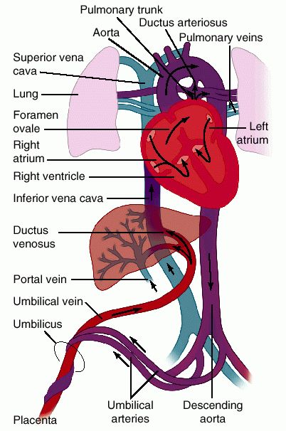
fetal circulation.
fetalcirculation3-1.gif


  • Gas exchange occurs in the uteroplacental circulation
  • fetal HG has a greater )2 affinity than adult HGb and oulls O2 from maternal blood
  • umblical arteries carry deoxygenated blood to placenta, umblical veing carry oxygenated from placenta to the portal system
  • changes following birth
  • lung expansion causes an increased pulmonary blood flow leading to an increase in relative blood oxygenation
  • a decreasing serum level of prostoglandin E results in ductus arteriosus closure; umblical cord clamping results in the end of placental circulation and increase in systemic vascular resistance
  • this increased vascular resistance, in trn induces the ductus venosus closure and umblical artery and vein constriction
  • left atrial pressure increases due to increased pulmonary blood flow, and umbilical circulation decreases, causing a decrease in inferior vena cava pressure.
  • decrease in inferior vena cava pressures leads to foramen ovale closure.
 
cardiac transplantation

  1. indicated for end stage cardiac disease (e.g. CAD, congenital DZ. cardiomyopathy with an estimated survival of <2 yrs
  2. contraindicated in pts. with pulmonary HTN. and Renal insufficiency. COPD, or other terminal diseases, smokers and pts > 70 yrs old are also excluded
  3. acute rejection is common
  4. most death occur in the initial 6 m after transplant, 5-yr survival is 70%
__________

case 1-
25 yr old man presents to PCP for a wellness checkup, he has been in good health. His only complain is recurrent Achilles tendon and hamstring pain that occurs following significant exertion. He has noticed some small bumps in the back of his heels and knees in the past yr in the regions of the pain. He denies medical conditions but states that his father died last year at a young age from a heart attack. He denies substance abuse, review of system and the remainder of his family is unremarkable. P.E is normal, but he has multiple small hard nodules behind his knees and heels.

Differential:
Chronic tendonitis, crystal arthropathy, hypercholesterolemia

labs unremarkable
ECG normal sinus
biopsy of the lesion shows a large collection of cholesterol laden material

DX. Familial hypercholesterolemia (hetrozygote)
RX

  1. the pt was placed on a regular exercise regimen and a low fat low cholesterol diet
  2. the pt was prescribed a regimen of simvastatin and ezetimibe
a repeat lipid analysis in 1 month found the LDL to be 155mg/dl
the tendon xanthomas gradually regressed
the pt was followed regularly to confirm an adequate reduction of lipid levels and cardiac screening.

The majority of cases of hypercholesterolemia are acquired

  1. steps to DX
  2. congenital hypercholesterolemia
  3. inherited form of hypercholesterolemia in which a genetic defect causes abnormally high levels of total cholesterol, LDL and/or triglycerides
common types:

  1. familial hypercholesterolemia autosomal dominant defect in LDL receptors with an associated increased total cholesterol and LDL : the dz is much more severe in homozygotes than hetrozygotes
  2. hypercholesterolemia and hypertriglycerdemia associated with an increased hepatic production of apolipoprotein B-100 protein
  3. Familial defective apolipoprotein B-100 similar to FH except that the defect is in the LDL particle and not LDL receptor
  4. history: tendonitis around xanthomas, xanthlesmas, cholesterol emboli are seen in the retina on fundoscopic exam
  5. total cholesterol and LDL >250 mg/dl in hetrozygotes and >600mg/dl in homozygotes
  6. triglycerides are elevate >200 in familial combined hyperlipidemia and a biopsy of xathomas will detect collections of cholesterol; genetic testing is available but typically uneeded for dx
  7. RX:
  8. healthy diet, exercise
  9. pt education
  10. lipid lowering agents , consisting of one statin and at least one other cholesterol lowering drug to achieve LDL levels <160 mg/dl or lower depending on cardiac risk factors
  11. trig drugs maybe added if required
Blood serum cholesterol levels should be collected from a fasting pt (12-14 hrs) to minimize post prandial influence.
 
[MEDIA]http://www.youtube.com/watch?v=8S8aZolpfCo&feature=related[/MEDIA]​
 
can't go wrong with a couple of aunty ji's teaching you about the EKG? :lol:..
 
[MEDIA]http://www.youtube.com/watch?v=A_xHXvWBNLU[/MEDIA]
[MEDIA]http://www.youtube.com/watch?v=HIB_F60THEU&feature=related[/MEDIA]
[MEDIA]http://www.youtube.com/watch?v=ex1k_MPF-w4[/MEDIA]
[MEDIA]http://www.youtube.com/watch?v=ecTM2O940mg&feature=related[/MEDIA]
[MEDIA]http://www.youtube.com/watch?v=Z5w8N6NBDys&feature=related[/MEDIA]
[MEDIA]http://www.youtube.com/watch?v=Rk6cWgNOVbk[/MEDIA]


Wow G.S. you seem to have put a lot of effort into these notes mashaAllah. I will inshaAllah take my time to go through these...it'll be good revision for me. (I'm a fresh graduate from med school)
JazakAllahu khairan!
 
[MEDIA]http://www.youtube.com/watch?v=_ITOH5LdAwg[/MEDIA]

JV catheter

indications:
Major surgery
drug admin- vasoactive drip meds.
hyperalimentation ( TPN)
chemo
prolonged abx therapy
CVP pressure monitoring
pulmonary artery cath.
transverse cardiac pacing
temporary hemodylaysis
Aspiration of air emboli

relative contraindications
previous carotid surgery
documented carotid disease
superior vena cava obstruction
severe trauma of the neck
coagulopathies


Jazakillah Khair sis Skye. Somehow I never noticed this thread until today. I will defo make use of it in the future. It looks really good MashaAllah.

May Allah reward you abundantly :wub:
 
Status
Not open for further replies.

Similar Threads

Back
Top