
You are using an out of date browser. It may not display this or other websites correctly.
You should upgrade or use an alternative browser.
You should upgrade or use an alternative browser.
Death in a scientific point of view and Islam point of view
- Thread starter lmaestro
- Start date
- Replies 28
- Views 11K
British Wholesales - Certified Wholesale Linen & Towels | Halal Food Gastronomy | PHP 8.4 patch for vBulletin 4.2.5
جوري
Soldier Through It!
- Messages
- 27,759
- Reaction score
- 6,095
- Gender
- Female
- Religion
- Islam
it means that the cells in your body have aged and can no longer do their function properly.. take any organ in the body and you'll see normal changes that happen with age.. Any organ.
the heart will lose its contractile ability as well as its elasticity, your glomerular filtration rate and renin-angiotensin system activity decreases with age, your cytochrome P450 system as well as other isozymed decrease with age.Your lung capacity and AP diameter goes down hill or rather I should say increases to resemble an emphysematous state.. I mean take any organ and you'll see a decrease in function, might not be considered 'pathological' but certainly considered change due to age..
No one has been able to find the fountain of youth. You can't get it out of a jar or plastic surgery-- tis man's lot.. Allah has decreed death for us as is stated in suret al- waqi3a ..
[media]http://www.youtube.com/watch?v=VlgC5CKh26U[/media]
hope that helped insha'Allah?
What do they mean by dying from natural causes/old age?
It's the inability of an organic body to regenerate or replenish its functional organs or parts.
Lobsters for some reason seem to be able to live forever, or at the very least they age so gracefully that we just don't find one that has died of natural causes. There are no mearsuable signs of decresing health or metabolism or reproductive abilities.
In most animals there is a genetic code which switches on the growth of certain organs and body parts, and conversely a genetic code which switches off these growing patterns. Most animals have these and it's quite apparent. fom the growth of your height, your limbs, your changes in metabolism, the changes in organ functionality, and these guide you through the complex stages of puberty as well.
If we can understand more about lobsters then perhaps one day humans could age so effortlessly that we may seem to live eternally.
جوري
Soldier Through It!
- Messages
- 27,759
- Reaction score
- 6,095
- Gender
- Female
- Religion
- Islam
Learn all you ever wanted to know about Lobsters here
Apparently nothing beats death not even cryopreservating...
He is the end event of all..
'natural' or otherwise..
cheers!
Apparently nothing beats death not even cryopreservating...
He is the end event of all..
'natural' or otherwise..
cheers!
Woodrow
May Allah have mercy on him رحمة الله عليه
- Messages
- 17,217
- Reaction score
- 4,224
- Gender
- Male
- Religion
- Islam
Death is a very natural part of the life process if our bodies are left to proceed with out intervention such as disease or accident. So, a natural death can be considered the fulfillment of bodily functions. Our bodies are so well designed that in our younger years much of our bodily functions are designed for replacing and rebuilding damaged tissue and organs. As we age this is no longer needed and our body begins a slow shutting down process, this process lasts long enough for us to prepare and accept the completion of our life. this is a natural death or so called dieing of old age.
Learn all you ever wanted to know about Lobsters here
Apparently nothing beats death not even cryopreservating...
He is the end event of all..
'natural' or otherwise..
cheers!
I'm sorry, I couldn't find the one that mentioned an end to the average life cycle, the only thing it said was that adulthood is reached in roughly 11 years. Which article were you looking at?
جوري
Soldier Through It!
- Messages
- 27,759
- Reaction score
- 6,095
- Gender
- Female
- Religion
- Islam
This one!
http://www.lobsters.org/ldoc/ldocpage.php?did=430
fromLobsters are long-lived animals. Their natural life expectancy is estimated to be somewhere between 50 and 100 years. The largest lobsters ever measured weighed in at more than 40 pounds!
http://www.lobsters.org/ldoc/ldocpage.php?did=430
جوري
Soldier Through It!
- Messages
- 27,759
- Reaction score
- 6,095
- Gender
- Female
- Religion
- Islam
Lobsters aside.. to the original poster, if you want to learn more about apoptosis (what is known as programmed cell death) here is an article which you won't find on the web and I can't link you to it.. but you may find various others on google, describing the same process...
simply as we have a life span so too do cells. It is as br. Woodrow stated a natural process.. except everything is truly a new rebirth... Just like you came from the darkness of the womb to experience glorious life, and just like you close your eyes in the darkness of night and find yourself in the gilded city of prague walking with your long lost friend.. so too in darkness and cold stench of death is there a rebirth into eternal life... so the way you should look at it, whether some horrible twist of fate cut life short, a disease or accident, or even through 'programmed' death as such we see in the cells.. it is all very natural..
simply as we have a life span so too do cells. It is as br. Woodrow stated a natural process.. except everything is truly a new rebirth... Just like you came from the darkness of the womb to experience glorious life, and just like you close your eyes in the darkness of night and find yourself in the gilded city of prague walking with your long lost friend.. so too in darkness and cold stench of death is there a rebirth into eternal life... so the way you should look at it, whether some horrible twist of fate cut life short, a disease or accident, or even through 'programmed' death as such we see in the cells.. it is all very natural..
Paul Anderson, MD, PhD
UpToDate performs a continuous review of over 375 journals and other resources. Updates are added as important new information is published. The literature review for version 15.1 is current through December 2006; this topic was last changed on April*18,*2006. The next version of UpToDate (15.2) will be released in June 2007.
INTRODUCTION — Apoptosis, or physiologic cell death, differs substantially from necrosis, or pathologic cell death. Unlike necrosis, apoptosis is essential for the following processes [1-3]: Morphogenesis during embryonic development Normal cell renewal Elimination of immune effector cells that proliferate in response to microbial infection.
The morphological features of apoptosis have been appreciated for decades: cytoplasmic and nuclear condensation, fragmentation of nuclei into membrane enclosed "apoptotic bodies," and surface expression of opsonic receptors that allow neighboring parenchymal cells to rapidly phagocytose and digest the corpse [4-6]. A key feature of this physiologic death process is the preservation of plasma membrane integrity. Rapid digestion of the contained apoptotic corpse avoids the recruitment of inflammatory cells which typically cause significant "collateral damage" to surrounding normal tissues.
A review of the relationship between apoptosis and autoimmune disease is presented here. Before discussing this interaction, it is helpful to briefly review the biology underlying apoptosis.
MOLECULAR MECHANISMS OF APOPTOSIS — The biochemical pathways responsible for apoptotic cell death have been elucidated in some detail. A genetic framework for the core death program has been provided by studies of a simple multicellular eukaryote, Caenorhabditis elegans [7].
Caenorhabditis elegans — Microscopic observations of C. elegans, a translucent nematode, have identified a subset of cells destined to die by apoptosis. Chemical mutagenesis has allowed the identification of genes that regulate this process.
In the scheme presented in Figure 1, ced-3 is a downstream effector of the apoptotic program (show figure 1). It is activated by ced-4, which is inhibited by ced-9. In turn, ced-9 is inhibited by egl-1. It follows from this scheme that the expression or activation of egl-1 results in apoptotic cell death.
Higher eukaryotes — Although the genetic blueprint utilized by C. elegans is followed in higher eukaryotes, the situation is complicated by the expression of multiple genes that correspond to each step delineated in the nematode: Just as ced-3 is a downstream effector of apoptosis in C. elegans, ced-3 orthologs comprise a family of proteases in higher eukaryotes that digest structural and enzymatic constituents of the cell to bring about apoptotic death [8]. The ced-4 orthologs (APAFs, for apoptosis activating factors) bind to the caspases and facilitate their conversion from inactive zymogens to active proteases [9]. The ced-9 and egl-1 orthologs comprise a large family of proteins related to bcl-2 [10], an oncogene that is transcriptionally activated by the t(14;18) chromosome translocation found in B-cell follicular lymphomas [11,12]. (See "Clinical and pathologic features of follicular lymphoma"). Overexpression of bcl-2 inhibits apoptosis in these cells, an essential component of lymphomatous growth. Just as ced-9 prevents cell death and egl-1 promotes cell death (by preventing the function of ced-9), individual members of the bcl-2 family can interact with one another to either promote or inhibit apoptosis [13].
**Triggering apoptosis — In higher eukaryotes, the core death program can be triggered from without (external or extrinsic) or from within (internal or intrinsic) the cell: External triggering involves the ligation of dedicated death receptors by soluble or cell-associated ligands [14]. Internal triggering occurs when cells respond to environmental stress (eg, heat, x-rays, ultraviolet irradiation) by altering the function of mitochondria, an organelle that is essential not only for cell survival, but also for regulating entry into cell death.
These extrinsic and intrinsic entryways into the death program are depicted schematically in Figure 2 (show figure 2). In this example, ligation of the dedicated death receptors CD95 (Fas) or TNF-RI, results in the recruitment of adaptor molecules (eg, FADD and TRADD) [9,15-17].
**Proteolytic cascade — The adaptor molecules subsequently bind to both the cytoplasmic domain of the dedicated death receptor and to the inactive form of caspase-8, an "upstream" caspase that initiates a proteolytic cascade leading to cell death. Because the zymogenic form of caspase-8 possesses low levels of protease activity, its aggregation allows one molecule to proteolytically activate an adjacent molecule, thereby initiating the proteolytic cascade. This culminates in the activation of effector caspases (eg, caspase-3) which directly cleave structural proteins (eg, nuclear lamins and cytoskeletal gelsolins [18]) and enzymes (eg, DNA-associated protein kinase, poly(ADP)ribose polymerase, etc.) to bring about apoptotic cell death.
In response to environmental stress, changes in the permeability of the outer mitochondrial membrane results in the release of cytochrome C and other effector proteins. Cytochrome C is a cofactor that allows APAF-1 to promote the activation of caspase-9, an "upstream" caspase that is essential for initiation of the intrinsic death program; the subsequent activation of effector proteases, including caspase-3, results in apoptotic cell death [19].
These basic pathways that lead to apoptosis are regulated by several proteins that bind to death receptors, adaptors, or caspases to modulate their function [20,21]. Examples include bcl-2 family members binding to APAFs, and inhibitors of apoptosis (IAPs) binding to caspases [22]. Not surprisingly, entry into apoptosis is a highly regulated process.
Noninflammatory phagocytosis — One of the hallmarks of apoptosis is that cells undergoing programmed death are normally phagocytosed by macrophages without activating an inflammatory or immune response. The mechanism for the efficient removal of apoptotic cells is not fully elucidated. The efficiency of the phagocytic process depends upon the presence of some normal components of plasma including complement components C1q, C3, C4, and members of the pentraxin family including pentraxin-3 (PTX3) C-reactive protein, and serum amyloid P component [23]. Binding of protein S, an antithrombotic plasma protein, to phosphatidylserine on the surface of apoptotic cells also promotes their phagocytosis [24].
The Long Island lobster deaths are a well documented case. If that is the basis for the person declaring "natural" life expectancy of the lobster to be 50-100 years then I don't know what to say to him/her. Those are figures that relate to the rise of the fishing industry in the early 19th century, (which is an external factor) and quite obviously the average life expectancy of lobsters dropped as they were being eaten. That is not to say that the natural life expectancy is the same.
All we know is that they are capable of living beyond a 100 years, and from some old reports 150 years, keeping in mind that we are speaking of natural causes and not external factors such as fishing or terminal illness.
David Foster Wallace (2005). Consider the Lobster and Other Essays. Little, Brown & Company. ISBN 0-31-615611-6.
More importantly, they do not experience the process of getting old with age. A 100 year old lobster can be just as healthy and functional as a 50 or 25 year old lobster and it is that key to retaining their youthful vitality as they age that scientists are interested in. That may well be the fountain of youth.
IbnAbdulHakim
IB Addict
- Messages
- 16,476
- Reaction score
- 2,628
- Gender
- Male
- Religion
- Islam
Lobsters for some reason seem to be able to live forever, or at the very least they age so gracefully that we just don't find one that has died of natural causes. There are no mearsuable signs of decresing health or metabolism or reproductive abilities..
now i cant help but wonder, how old is the oldest lobster????
now i cant help but wonder, how old is the oldest lobster????
:salamext:
The World's Oldest Lobster- 110 million years old
Source
IbnAbdulHakim
IB Addict
- Messages
- 16,476
- Reaction score
- 2,628
- Gender
- Male
- Religion
- Islam
it says old fossilised lobster, so its dead? did it actually live and breathe for 110 million years? :O
IbnAbdulHakim
IB Addict
- Messages
- 16,476
- Reaction score
- 2,628
- Gender
- Male
- Religion
- Islam
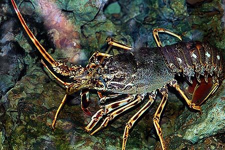
it looks sooooooooooooo good though !! its like some golden ornament, seriously i was in awe when i saw the picture of that lobster (the colored one)
IbnAbdulHakim
IB Addict
- Messages
- 16,476
- Reaction score
- 2,628
- Gender
- Male
- Religion
- Islam
this one:
IbnAbdulHakim
IB Addict
- Messages
- 16,476
- Reaction score
- 2,628
- Gender
- Male
- Religion
- Islam
:salamext:
Nyccccccccccccccccccc! How old is that? I mean if the fossil thingy u take that out, then is it still 110 million years old?
didnt i ask you that? :heated:
Woodrow
May Allah have mercy on him رحمة الله عليه
- Messages
- 17,217
- Reaction score
- 4,224
- Gender
- Male
- Religion
- Islam
it says old fossilised lobster, so its dead? did it actually live and breathe for 110 million years? :O
The article says it is the fossil of a juvenile lobster, so it was probably less than 3 years old.
But, returning to topic it does seem that death is actually a gift and for a purpose.
For those that believe that life is the result of spontaneous evolution, I often wonder how they explain death. There is no scientific reason anything should die. In any organism all of the living cells are replaced constantly. At any given moment us humans do not have any living cells that are over 7 years old, with the exception of the nervous system and nerve cells do not regenerate, nor appear to age, except through injury or disease. Natural death of the nervous system does not occur until after the death of the supporting cells and organs. There does not seem to be any basis as to why death should occur. we can explain the actual process, but there does not seem to be any reason why the process is even there.
Unless you want to entertain the possibility that the body is simply a temporary vehicle for the development(education?) of something else, and once that development is complete the body is no longer needed.

Similar Threads
Add to Homescreen
How to install the app on iOS
Follow along with the video below to see how to install our site as a web app on your home screen.
Note: This feature may not be available in some browsers.